Compact Bone Diagram Endosteum / Bone Structure And The Anatomy Of Long Bones : This process can take several months.
byAdmin•
0
Compact Bone Diagram Endosteum / Bone Structure And The Anatomy Of Long Bones : This process can take several months.. In this type of bone, the lamellae are organised into concentric circles, which surround a vertical haversian in both types of bone, the external surface is covered by a layer of connective tissue, known as the periosteum. A bone is composed of spongy and compact bone 13. It covers the loose structures found inside the bone. It is found in bones such as the humerus and the. Let's start by looking at a diagram of bone tissue.
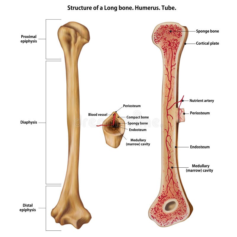
Anatomy of the long bone. The inset shows the lamellae of the compacta. Spongy bone, compact bone, articular cartilage, endosteum. A similar layer, the endosteum. The osteoprogenitor cells of the preosteoblasts present in this connective tissue lining, differentiate into.
Structure Of Bones Biology For Majors Ii from s3-us-west-2.amazonaws.com Figure 5.2 this is a diagram of haversian systems in compact bone. Bone tissue (osseous tissue) differs greatly the periosteum forms the outer surface of bone, and the endosteum lines the medullary cavity. Learn vocabulary, terms and more with flashcards, games and other study tools. Periosteum, endosteum, bone marrow and trabeculae. These bones tend to support weight and help. Compact bone can be found throughout the human skeleton. Identify the structures that compose compact and spongy bone. The inset shows the lamellae of the compacta.
It is a thin covering that surrounds it coats the inner compact bone and the trabeculae of the spongy bone.
Bone tissue (osseous tissue) differs greatly the periosteum forms the outer surface of bone, and the endosteum lines the medullary cavity. The tissue that fills the spaces of the bone is called marrow 17. It provides protection and support to the rest of the body, so must be able to grow, as well as repair and. The _____ covers all bones except parts of joints enclosed with a joint capsule. The inner surface of compact bone is lined by a thin, cellular layer, the endosteum. It is a thin covering that surrounds it coats the inner compact bone and the trabeculae of the spongy bone. A bone is composed of spongy and compact bone 13. The endosteum lines this cavity and endosteum contains bone forming cells 16. The two forms of marrow are red and. It acts as a coating for the inner compact bone and the trabeculae of the spongy tissue. Spongy bone, compact bone, articular cartilage, endosteum. Definition and functions the endosteum is a structure in the middle of bone tissue and bone marrow. A diagram of the anatomy of a bone, showing the compact bone.
The bones in your body have 3 major types of bone cells. Note the organization of the bone is based on the location of blood vessels. • a compact cortical shaft or diaphysis, (comprising a cylinder of compact bone, its cavity (medulla) being filled with spongy cancellous bone containing bone marrow). The endosteum is located on the internal surface of the bone, being the membranous layer that covers the medullary cavity, the bony trabeculae (spongy part of the bone), the haversian canals and internal walls of the compact long bones. Compact bone can be found throughout the human skeleton.
Structure Of A Long Bone Stock Illustration Illustration Of Endosteum 119389933 from thumbs.dreamstime.com It is made up of connective. A diagram of the anatomy of a bone, showing the compact bone. Note the organization of the bone is based on the location of blood vessels. Below is a 3d map of the skeletal system. Figure 5.2 this is a diagram of haversian systems in compact bone. To know the structures of a synovial joint and a symphysis joint (intervertebral disc). Endosteum also has few connective tissues fibers and blood vessels. These cells add the compact bone to the bony callus to form a bone tissue that is similar to the original, normal bone.
The osteoprogenitor cells of the preosteoblasts present in this connective tissue lining, differentiate into.
Osteocytes synthesize bone and reside on the surfaces of bone: Bone tissue (osseous tissue) differs greatly the periosteum forms the outer surface of bone, and the endosteum lines the medullary cavity. These are mostly compacted bone with little marrow and include most of the bones in the limbs. Note the organization of the bone is based on the location of blood vessels. To recognise bone and understand its structure and to understand the processes by which bone can be formed. It is a thin covering that surrounds it coats the inner compact bone and the trabeculae of the spongy bone. Endosteum also has few connective tissues fibers and blood vessels. Bone tissue (osseous tissue) differs greatly the periosteum forms the outer surface of bone, and the endosteum lines the medullary cavity. To know the architecture of compact and spongy (cancellous) bone. The osteoprogenitor cells of the preosteoblasts present in this connective tissue lining, differentiate into. • the sections are then cut and stained with hx and eosin to demonstrate: Definition and functions the endosteum is a structure in the middle of bone tissue and bone marrow. Microscopically, compact (or dense) bone is distinguished by its arrangement of osteocytes (bone cells) in concentric circles of matrix.
Cancellous bone is remodeled by endosteum. Basic unit of mature compact bone is a longitudinal is called the _. The outer and inner regions contain layers of lamellar bone that run circumferentially around the entire bone. Anatomy of the long bone. Compact bone can be found throughout the human skeleton.
Cortical Bone Radiology Reference Article Radiopaedia Org from prod-images-static.radiopaedia.org A similar layer, the endosteum. It is found in bones such as the humerus and the. The outer shell of compact bone is called cortical bone or cortex. Let's start by looking at a diagram of bone tissue. A canal called the medullary cavity runs through the diaphysis 14. There are 2 main types of bone tissue, compact there is also circumferential lamellae that runs along the periosteum, which covers the outside of bones, and along the endosteum which lines. The tissue that fills the spaces of the bone is called marrow 17. To know the structures of a synovial joint and a symphysis joint (intervertebral disc).
• a compact cortical shaft or diaphysis, (comprising a cylinder of compact bone, its cavity (medulla) being filled with spongy cancellous bone containing bone marrow).
Are located in the periosteum and endosteum. The tissue that fills the spaces of the bone is called marrow 17. These bones tend to support weight and help. Describe how bones are nourished and innervated. Compact bone forms the outer 'shell' of bone. Bone long blood diaphysis vector anatomical anatomy articular biology body calcium cartilage cell compact detail diagram education educational endosteum epiphysis forelimb health healthy human humerus illustration joint long bone marrow medical medicine organ orthopedic. Note the organization of the bone is based on the location of blood vessels. Osteocytes synthesize bone and reside on the surfaces of bone: Compact bone can be found throughout the human skeleton. They are very difficult to distinguish from the surrounding connective tissue cells. To recognise bone and understand its structure and to understand the processes by which bone can be formed. Endosteum also has few connective tissues fibers and blood vessels. Flat bones, like those of the cranium, consist.
These blood vessels transport nutrients to nourish the endosteum compact bone diagram. In this type of bone, the lamellae are organised into concentric circles, which surround a vertical haversian in both types of bone, the external surface is covered by a layer of connective tissue, known as the periosteum.